We’re all wondering what the future of
#spatialtranscriptomics (ST) has in store. Well, I think the near future will look a lot like the new 6000-plex dataset reviewed here (link to the original source at the end of this post).
SAMPLE
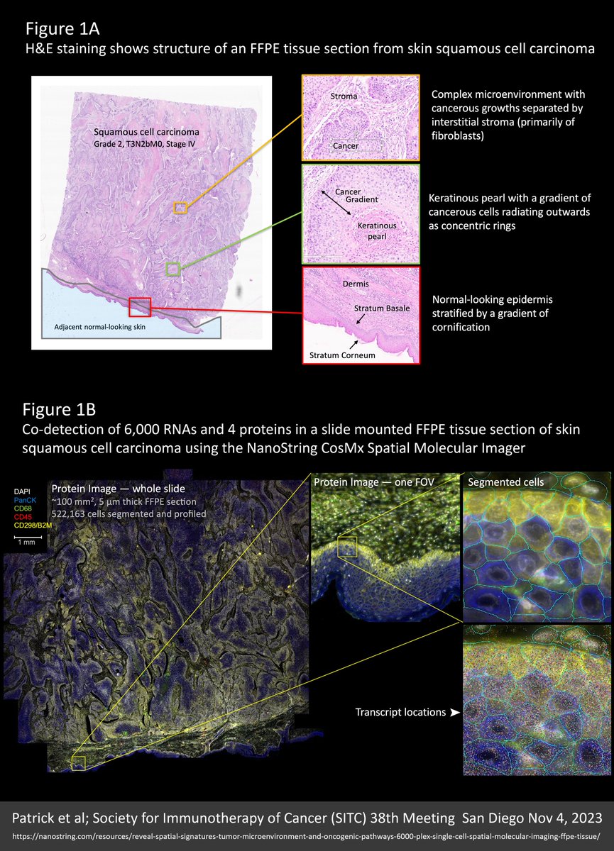
An entire 100 mm² intact 5 µm FFPE section from a skin squamous cell carcinoma was profiled for 6,000 unique RNA targets plus four protein markers (CD298/B2M, CD45, PanCK, CD68) and DAPI for nuclear staining using the NanoString CosMx Spatial Molecular Imager. Altogether, over half a million cells were profiled, segmented, and analysed from billions of mapped molecule coordinates.
With this particular cancer sample, the skin surface looked normal but the underlying tumour structure was complex. See Figure 1A for imagery and further detail.
Note in Figure 1B how protein markers are critical to defining accurate cell boundaries for segmentation and plotted transcripts underscore the super density of this dataset.
OPTICAL CROWDING
A common pain-point in the ongoing debate over the relative merits of the $NSTG NanoString CosMx and $TXG 10X Genomics Xenium molecule-mapping ST platforms, is the anguish in avoiding “optical crowding” and saturating the “signal space” with either too many targets, higher expressing targets, or both. I only discovered these terms following 10x Xenium discussions; they were never part of the NanoString lexicon. With this dataset we see why: Most of the cells surveyed contained over 2,500 transcripts and some mapped up to 5,000 transcripts per cell, yet there is no evidence of overcrowding. What’s more, these very high counts were obtained from accurately segmented cells that are typically 300-400% smaller than you obtain when similar cells are segmented using the Xenium “expansion” method, detailed in my recent X post reviewing data from the lab of Dr Yutaka Suzuki at University of Tokyo. Figure 2 from that previous X post is reproduced below. This only makes this 6000-plex dataset 300-400% more impressive! See Figure 3A for transcript density plots cor this skin carcinoma sample.
NEGATIVE CONTROLS
Another discussion point is negative controls and the smoke and mirrors of “false discovery”. Note that negative controls vary across the tissue relative to the density of the cellular matrix (Figure 3A). This is simply because individual molecules are “sticky” and the more cellular matrix available the more get stuck, such that a distribution plot of negative control counts/cell (in Figure 3A) recapitulates a detailed image of the tissue structure. That said, counts are very low (less than a total of 4 per cell from the 10 separate negative controls included in the assay).
CELL TYPING
A UMAP dimensional reduction plot of cell types shows a predominance of fibroblasts, keratinocytes, and macrophages. On the cancer side, a distinctive “keratinous pearl” cell type and at least eight separate but interrelated cancer subtypes are evident (Figure 3B). A niche analysis beautifully resolves seven intricately detailed spatial population clusters, emphasizing the complexity of this carcinoma (Figure 3B).
CELL TYPE VERIFICATION
Super density ST data (thousands of targets and thousands of transcripts per cell) allows for greater certainty and nuance in cell typing. This is underlined by the multitude of cancer type variants and the fine gradation between them. Accurate cell typing was confirmed by how well the spatial distribution of cell types corresponded with orthogonal data from H&E images, marker genes that most differentiate groups of cells, and alignment with protein stains. Figure 4A demonstrates colocalization of cell types, marker genes, and stained proteins for three major cell types.
NEW BIOLOGY
Interestingly, a gradation of cancer cell types was found to radiate in clearly defined rings around keratinous pearl cell core structures. These cancer subpopulations resolved from cells that lack any defining marker gene (Figure 4B).
CONCLUSIONS
Unsurprisingly, higher plex assays generate more biological insight and greater spatial detail. Kilo-plex assays will be an expectation.
Assays need a protein information layer. This also will be an expectation. Not only is this critical for accurate cell segmentation (where inaccuracies contribute the most noise and obfuscation of results) but also for biological insight and orthogonal multiomic validation. The trend will be to higher and higher plex same-cell protein data that digitally overlays the RNA layer.
I’m so very excited by this new wave of spatial transcriptomics (ST) technologies. They will not only massively expand biological insight but also our appreciation for the exquisite beauty of multicellular organisation.
Find the original Poster publication for this NanoString 6000-plex dataset here:
@AlbertVilella @BlauerPlums @lee_spraggon @NeBanovich @ArpitaBKulkarni @GenomicsCow @aruthak @GESTALT_sp @HumphreysLab @DavidPCook @DrJasPlummer @TheGregoryLab @m_mohenska @kirkbjensen @nikhil_seq @Kellieiswise @pinkney_holly @ioavlachos @rkhyip @LGMartelotto