EHJ-IMP Editor-in-Chief✓

@EHJIMPEiC

#EHJIMP Editor-in-Chief X account. Posts by @alessia_gimelli and her team. #EACVI #cvimaging #echofirst #CVNuclear #yesCCT #whyCMR

336 Following1.7K Followers1.5K Posts
  1. EHJ-IMP Editor-in-Chief✓@EHJIMPEiC
    EHJ-IMP Editor-in-Chief
    RT @EHJIMPEiC: Hot off the press #EHJIMP🔥🔥 🤖 Automated Echocardiographic Assessment of Dilated Cardiomyopathy Using Deep Learning in a Resource-Limited Setting 🤖 📌 Key Findings: Population: 186 patients with dilated cardiomyopathy (DCM) at Jakaya Kikwete Cardiac Institute (JKCI), Tanzania Approach: U-Net deep learning framework for automated left ventricular (LV) segmentation and ejection fraction (EF) estimation, using models developed on CAMUS (still images) and EchoNet-Dynamic (video) Domain shift: The CAMUS-trained still-image model produced fragmented predictions on JKCI data, unsuitable for LV volume estimation EF estimation after fine-tuning (EchoNet-Dynamic): – MAE: 7.21% → 4.63% – RMSE: 9.84% → 6.66% LV segmentation (Dice similarity coefficient): – End-systole: 0.90 – End-diastole: 0.84 🔗 Read the full article: @VikashJaiswalMD @EHJIMPEiC @JGrapsa @ESC_Journals #Echocardiography #DeepLearning #DilatedCardiomyopathy #CardiovascularImaging #GlobalHealth #EHJIMP
    01101.7K
  2. EHJ-IMP Editor-in-Chief✓@EHJIMPEiC
    A new study published in the #EHJIMP highlights the potential of deep learning-based echocardiography to enhance prognostic assessment in patients with heart failure with reduced ejection fraction (HFrEF). 🔬 Key findings The prospective observational study included 568 patients with newly diagnosed HFrEF, with a median follow-up of 4.8 years. 📊 Important results: 🔹 Ventricular arrhythmias: 49 patients (9%) experienced ventricular arrhythmias during follow-up, while 118 (21%) died. 🔹 Automated imaging biomarkers: Deep learning was used to measure global longitudinal strain (GLS) and mechanical dispersion (MD), with optimal cut-offs of 9% and 70 ms, respectively. 🔹 Combined risk assessment: Patients with both impaired GLS and increased MD had a substantially higher risk of ventricular arrhythmias than patients with only one of these abnormalities: • Both abnormalities: HR 6.98 (95% CI 3.23–15.1) • Either abnormality alone: HR 2.84 (95% CI 1.38–5.86) 🔹 Incremental prognostic value: GLS and MD were independently associated with ventricular arrhythmias and showed potential additional prognostic value beyond left ventricular ejection fraction (LVEF). 🔹 Rapid, scalable analysis: Automated measurements were feasible in 96% of cases, with a mean processing time of just 6.4 ± 2.5 seconds per echocardiogram. 🔗 Read the full article: @VikashJaiswalMD @JGrapsa @EHJIMPEiC #HeartFailure #HFrEF #ArtificialIntelligence #DeepLearning #Echocardiography #GlobalLongitudinalStrain #MechanicalDispersion #VentricularArrhythmia #SuddenCardiacDeath #CardiacImaging #DigitalHealth
    Download
    110201K
  3. EHJ-IMP Editor-in-Chief✓@EHJIMPEiC
    Papillary fibroelastoma is a rare primary cardiac tumour with benign histology. Despite its benign nature, it can lead to serious complications, including thromboembolism, stroke, myocardial infarction, and dynamic coronary obstruction. This review at #EHJ-IMP highlights how multimodality imaging supports diagnosis and clinical management: 🔹 Transthoracic echocardiography (TTE) serves as an important initial imaging test. 🔹 Transoesophageal echocardiography (TEE) provides improved spatial and temporal resolution, helping define tumour mobility, attachment, stalk morphology, and relationships with nearby structures. 🔹 Cardiac CT offers detailed anatomical information and helps assess the tumour’s relationship with the cardiac valves, coronary arteries, and surrounding anatomy. 🔹 Cardiac MRI contributes to tissue characterisation and may help differentiate papillary fibroelastoma from other cardiac masses. 🔗 @VikashJaiswalMD @EHJIMPEiC @JGrapsa #EHJImaging #Cardiology #CardiacImaging #MultimodalityImaging #Echocardiography #CardiacCT #CardiacMRI #PapillaryFibroelastoma #CardioEd #CardioTwitter.
    Download
    019362.9K
  4. EHJ-IMP Editor-in-Chief✓@EHJIMPEiC
    A multicentre study published in European Heart Journal – Imaging Methods and Practice evaluated the relationship between ventricular fibrosis and recovery of LV function after catheter ablation for atrial fibrillation. The study included 134 patients with AF and LV systolic dysfunction. Following ablation, 101 patients (75.4%) achieved meaningful LV recovery. 🔹 Ventricular late gadolinium enhancement (LGE) was more common among patients without recovery: 69.7% vs 46.5%. 🔹 Patients who did not recover had nearly twice the LGE burden compared with responders: 6.6% vs 3.3% (p<0.001). 🔹 Each 1% increase in LGE burden was associated with a lower likelihood of LV recovery (RR 0.96; 95% CI 0.92–0.99; p=0.036). 🔹 The visual pattern and distribution of LGE were not associated with recovery. 🔗 @EHJIMPEiC @VikashJaiswalMD #AtrialFibrillation #CatheterAblation #CardiacMRI #LateGadoliniumEnhancement #HeartFailure #LeftVentricularFunction #MyocardialFibrosis #Cardiology #CardiacImaging #RhythmControl #Electrophysiology #CardioEd #CardioX
    Download
    0714662
  5. EHJ-IMP Editor-in-Chief✓@EHJIMPEiC
    A new report in #EHJIMP highlights a rare but potentially life-threatening complication of mitral transcatheter edge-to-edge repair (M-TEER): right-to-left shunting through an iatrogenic atrial septal defect (iASD). 🔍 Key findings from 444 patients undergoing successful M-TEER: ▪️ 6 patients (1.3%) required emergency iASD closure for acute deoxygenation. ▪️ All had severe tricuspid regurgitation, right ventricular (RV) dilatation, and impaired RV function. ▪️ In 5 of 6 patients, right atrial pressure exceeded left atrial pressure after M-TEER. ▪️ RV function was substantially impaired, with lower TAPSE (15 vs. 19 mm), fractional area change (27% vs. 43%), and RV free-wall strain (−13% vs. −22%). ▪️ Echocardiography underestimated invasively measured systolic pulmonary artery pressure (25.8 vs. 40.3 mmHg; P = 0.035). ▪️ Oxygen saturation decreased by 3–8%; one patient developed cardiogenic shock. Read the article: @VikashJaiswalMD @EHJIMPEiC #Cardiology #StructuralHeart #MTEER #MitralRegurgitation #Echocardiography #RightVentricle #CardiacImaging #Cardiotwitter
    Download
    28151.2K
  6. EHJ-IMP Editor-in-Chief✓@EHJIMPEiC
    Can clinically manifest cardiac sarcoidosis occur in the absence of LGE on CMR? A new study published in #EHJ-IMP provides evidence that clinically significant, metabolically active cardiac sarcoidosis may occur despite the absence of late gadolinium enhancement (LGE) on cardiac magnetic resonance (CMR). 🔬 Key findings • Among 46 treatment-naïve patients with clinically manifest, FDG-avid cardiac sarcoidosis, 4 (8.7%) had no detectable LGE on CMR. • All 4 LGE-negative patients presented with advanced conduction system disease; 3 had complete heart block. • Left ventricular ejection fraction was higher in LGE-negative than in LGE-positive patients (70% vs. 50%; P = 0.010). • Myocardial inflammatory burden, assessed by LV SUVmax on FDG-PET, was lower in LGE-negative patients (3.9 ± 0.9 vs. 9.6 ± 5.2; P = 0.037). • Three of the 4 LGE-negative patients received immunosuppressive therapy. 📄 Read the full article: @EHJIMPEiC @VikashJaiswalMD 🗣️#CardioTwitter #Cardiology #CardiacSarcoidosis #Sarcoidosis #WhyCMR #CVImaging #NuclearCardiology #FDGPET #EPeeps #HeartBlock #Arrhythmia #HeartFailure #MedEd #EHJIMP
    Download
    0915782
  7. EHJ-IMP Editor-in-Chief✓@EHJIMPEiC
    A new systematic review and meta-analysis in the #EHJIMP compared transthoracic echocardiography (TTE) with cardiac magnetic resonance (CMR) for the detection of left ventricular thrombus. The analysis included 18 studies involving 3,478 participants. Key findings: ✅ CMR sensitivity: 70.5% ✅ TTE sensitivity: 31.8% ✅ CMR specificity: 99.2% ✅ TTE specificity: 96.3% ✅ CMR AUC: 0.985 ✅ TTE AUC: 0.59 CMR demonstrated a clear diagnostic advantage, particularly for detecting thrombi that may be missed by standard echocardiography, including small or mural thrombi. However, the findings require careful interpretation. Most studies used late gadolinium enhancement CMR as the reference standard, and only five studies provided direct head-to-head comparisons using an independent reference standard. Echocardiographic protocols also varied, and the use of contrast-enhanced TTE was not consistent. The clinical message: ❤️ TTE remains an accessible and valuable first-line test. ❤️ CMR should be considered when TTE is negative but clinical suspicion remains high, when image quality is limited, or in patients at high risk for left ventricular thrombus. ❤️ Contemporary studies using standardized imaging protocols and independent validation are still needed. 🔗 Read the full study: @EHJIMPEiC @VikashJaiswalMD #LeftVentricularThrombus #CardiacMRI #CMR #Echocardiography #TTE #CardiacImaging #MyocardialInfarction #StrokePrevention #Cardiology #ImagingInnovation #CardiovascularMedicine
    Download
    213191.5K
  8. EHJ-IMP Editor-in-Chief✓@EHJIMPEiC
    A new study in the European Heart Journal – Imaging Methods and Practice evaluated whether measuring diastolic flow reversal in the distal abdominal aorta could improve the echocardiographic assessment of aortic regurgitation. Abdominal aortic holodiastolic flow reversal is an established marker of significant aortic regurgitation, but guidelines do not clearly define the optimal measurement location. The study evaluated 103 patients with moderate or severe chronic aortic regurgitation. Key findings: ✅ Distal flow-reversal assessment was feasible in 94% of patients ✅ Proximal assessment was feasible in only 54% ✅ Diagnostic yield was higher with distal measurements: 82% vs 50% ✅ In paired analyses, distal assessment showed 92% sensitivity, 86% specificity, and 88% accuracy for severe aortic regurgitation ✅ Reproducibility between and within observers was excellent ✅ In a small exploratory subgroup, distal echocardiographic findings showed 92% agreement with cardiac MRI 🔗 Read the full study: @EHJIMPEiC @VikashJaiswalMD @JGrapsa #AorticRegurgitation #AorticValveDisease #Echocardiography #CardiacImaging #CardiacMRI #ValvularHeartDisease #HeartValveDisease #Cardiology #ImagingInnovation #MedicalResearch #StructuralHeartDisease #CardioX #CardioEd
    Download
    120474.2K
  9. EHJ-IMP Editor-in-Chief✓@EHJIMPEiC
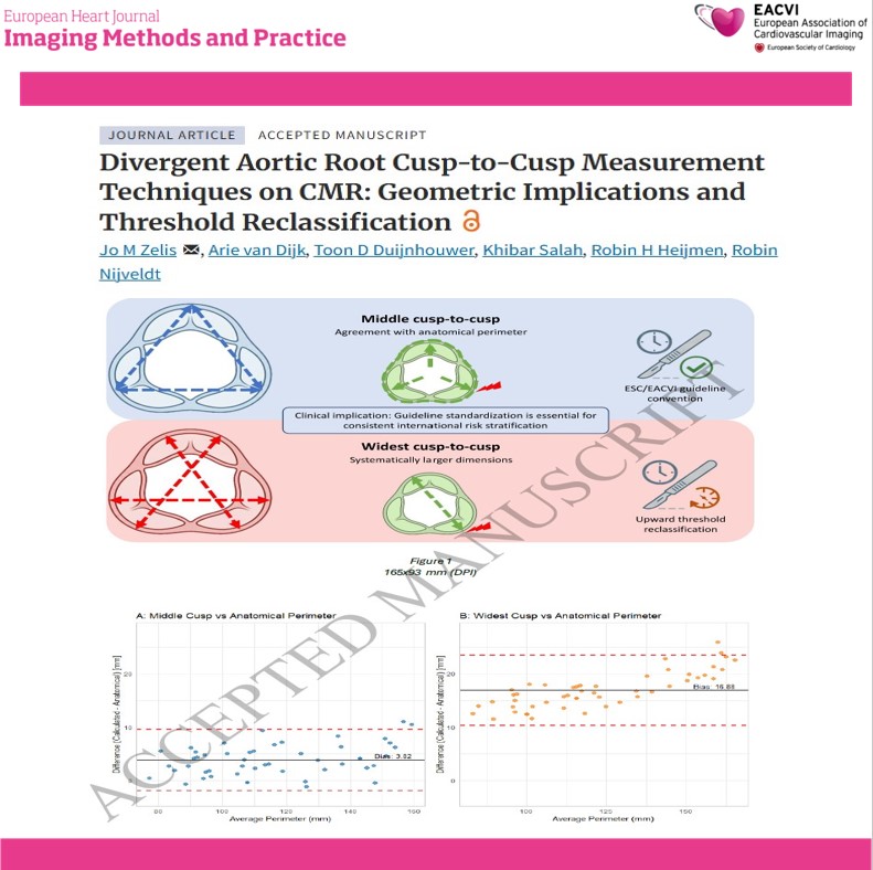
    A new study in the #EHJIMP examined differences between ESC and EACTS cusp-to-cusp measurement techniques using 3D cardiovascular magnetic resonance imaging. The study included 50 patients with a mean age of 42 years and compared: 🔹 Middle cusp-to-cusp measurements 🔹 Widest cusp-to-cusp measurements 🔹 Traced anatomical aortic root perimeter Key findings: ✅ Widest measurements were, on average, 3.60 mm larger than middle measurements ✅ Middle measurements showed better agreement with the traced aortic root perimeter ✅ Lin’s concordance correlation coefficient: 0.98 vs 0.80 ✅ Mean absolute error: 3.96 mm vs 16.88 mm ⚠️ Use of the widest measurement reclassified 10% of patients at the 40-mm threshold and 16% at the 45-mm threshold These findings highlight an important challenge in aortic surveillance: different guideline-recommended measurement conventions can place the same patient in different clinical risk categories. The study emphasizes the need for: 📌 Standardized aortic root measurement techniques 📌 Improved comparability across imaging studies 📌 Consistent international risk stratification 📌 Prospective validation against clinical outcomes 🔗 Read the full study: @EHJIMPEiC @VikashJaiswalMD @saramoscatelli7 @JGrapsa #AorticDisease #AorticRoot #CardiacMRI #CMR #CardiovascularImaging #AorticAneurysm #AorticSurgery #Cardiology #CardioTwitter #CardioX #CardioED
    Download
    01214948
  10. EHJ-IMP Editor-in-Chief✓@EHJIMPEiC
    EHJ-IMP Editor-in-Chief
    RT @EHJIMPEiC: A new state-of-the-art review in the #EHJ-IMP highlights how multimodality imaging can guide the complete management pathway for tricuspid regurgitation—from referral to surgery, transcatheter intervention, and follow-up. The review outlines the complementary role of: 🔹 Transthoracic echocardiography: initial assessment and disease severity 🔹 3D transesophageal echocardiography: detailed leaflet and annular anatomy 🔹 Cardiac magnetic resonance: reproducible assessment of right ventricular volumes and function 🔹 Cardiac CT: annular geometry, adjacent structures, device leads, and vascular access The authors emphasize that treatment decisions should integrate: ✅ Mechanism of tricuspid regurgitation ✅ Annular dilatation and leaflet tethering ✅ Right ventricular function and reserve ✅ Pulmonary hemodynamics ✅ End-organ involvement ✅ Surgical, transcatheter, or surveillance options 🔗 Read the full review: @EHJIMPEiC @VikashJaiswalMD #TricuspidRegurgitation #HeartValveDisease #StructuralHeartDisease #Echocardiography #CardiacMRI #CardiacCT #MultimodalityImaging #CardiacSurgery #TranscatheterTherapy #RightHeartFailure #HeartTeam #Cardiology #CardiovascularImaging #MedicalResearch #PatientCare
    080930
  11. EHJ-IMP Editor-in-Chief✓@EHJIMPEiC
    A new short report in the #EHJ-IMP highlights a rare but clinically important complication following mitral transcatheter edge-to-edge repair (M-TEER). Acute oxygen desaturation after M-TEER should prompt immediate assessment of: 🔍 Right ventricular function 🔍 Right atrial and pulmonary pressures 🔍 Residual or iatrogenic atrial septal defect 🔍 Direction of interatrial blood flow 🔍 New right-to-left shunting 🔍 Changes in left atrial pressure after repair Although an iatrogenic atrial septal defect is common after transseptal procedures and is often hemodynamically tolerated, elevated right-sided pressures or acute right ventricular dysfunction can reverse the direction of flow—resulting in a clinically significant right-to-left shunt and sudden hypoxemia. This report reinforces the importance of: ✅ Continuous hemodynamic monitoring ✅ Prompt transesophageal echocardiographic assessment ✅ Careful evaluation of right-sided heart function ✅ Identifying the mechanism of hypoxemia before considering device closure ✅ Multidisciplinary decision-making during structural heart procedures 🔗 Read the full report: @EHJIMPEiC @VikashJaiswalMD @saramoscatelli7 @JGrapsa #MTEER #MitralRegurgitation #StructuralHeartDisease #InterventionalCardiology #Echocardiography #TransesophagealEcho #RightHeartFailure #CardiacImaging #StructuralHeartInterventions #Cardiology #CardioX #CardioEd
    Download
    014281.7K
  12. EHJ-IMP Editor-in-Chief✓@EHJIMPEiC
    A recent study from the Yorkshire Heart Centre demonstrates how targeted CMR protocol redesign can reduce scan times and expand imaging capacity. The study evaluated a “Short Aorta” CMR protocol for patients referred primarily for thoracic aortic assessment. Unlike the standard protocol, the shorter approach focused only on the thoracic aorta when additional assessment of the aortic valve and left ventricular function was not clinically required. Key findings: ✅ 137 referrals were reviewed ✅ 69% were clinically suitable for the shorter protocol ✅ 46% of scans used the “Short Aorta” protocol after implementation ✅ Median scan time decreased from 37 to 18 minutes ✅ Approximately 1.2 hours of scanner time per week was saved ✅ With optimized scheduling, potential savings increased to 1.9 hours per week These findings highlight how thoughtful protocol redesign—not only new technology—can improve CMR workflow, reduce patient waiting times, and increase imaging capacity. The shorter protocol should be used selectively and with appropriate clinical triage. Patients requiring assessment of the aortic valve, left ventricular function, or other cardiac structures should continue to undergo the full CMR protocol. 🔗 Read the full study: @EHJIMPEiC @VikashJaiswalMD @saramoscatelli7 #CardiacMRI #CMR #CardiovascularImaging #AorticDisease #MedicalImaging #HealthcareInnovation #QualityImprovement #Radiology #Cardiology #ImagingEfficiency #PatientCare #CardioX #CardioEd
    Download
    189580
  13. EHJ-IMP Editor-in-Chief✓@EHJIMPEiC
    A new study in the #EHJ-IMP assessed cardiac image quality from single-probe handheld ultrasound devices. Authors evaluated 518 cine-loops from 206 patients using Butterfly IQ+, Butterfly IQ3, and Vave devices. Key findings: ✅ 88% of cine-loops were interpretable ✅ 91% of patients had at least one interpretable image ⚠️ Only 12% achieved good-quality imaging 📉 Image quality declined with increasing BMI ⚖️ No significant difference was observed by sex 🫀 Parasternal long-axis views provided the best image quality The findings support the potential of handheld ultrasound to assist with early detection of structural heart disease and point-of-care cardiac assessment. However, image quality assessment remains essential. Handheld ultrasound should complement not replace comprehensive echocardiography, particularly when evaluating complex valvular disease, cardiomyopathies, or Doppler-dependent conditions. The next step: integrating standardized image-quality assessment and artificial intelligence to identify inadequate studies and guide referral for formal echocardiography. 🔗 Read the full study: @VikashJaiswalMD @saramoscatelli7 @EHJIMPEiC #Cardiology #Echocardiography #POCUS #HandheldUltrasound #CardiacImaging #StructuralHeartDisease #MedicalResearch #CardiovascularMedicine
    Download
    136496
  14. EHJ-IMP Editor-in-Chief✓@EHJIMPEiC
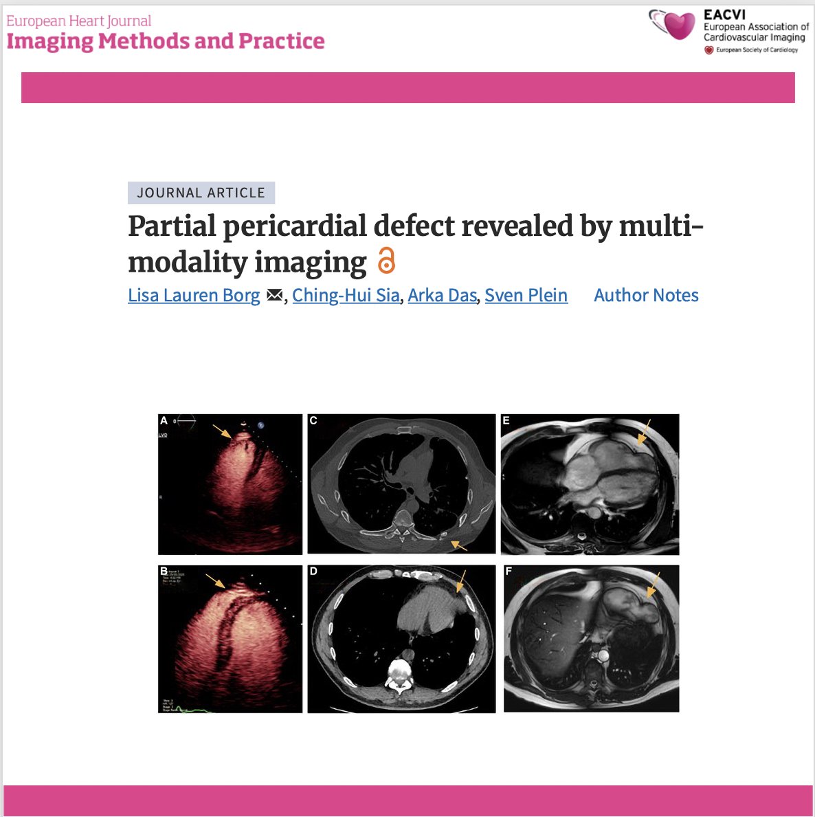
    🫀 Could a traumatic partial pericardial defect be hiding in plain sight? A fascinating multimodality imaging case highlights how echocardiography, CT and cardiac MRI uncovered an acquired partial pericardial defect with right ventricular apical herniation in a patient presenting with intermittent chest pain years after major thoracic trauma. 🔍 Key findings: ✅ Stress echocardiography showed an apparently dilated right ventricle with an aneurysmal-appearing apex, raising suspicion for structural heart disease. ✅ Chest CT demonstrated loss of the pericardial contour at the RV apex in the region of previous left-sided rib fractures. ✅ Cardiac MRI confirmed a partial pericardial defect with herniation of the RV apex, while showing preserved RV size, systolic function and normal myocardial tissue characteristics. ✅ As the patient remained clinically stable without evidence of progressive herniation or ventricular dysfunction, conservative management with serial imaging was chosen instead of surgery. 💡 Clinical takeaway: Not every "RV aneurysm" is myocardial disease. Multimodality imaging is essential to distinguish pericardial defects from cardiomyopathies and can reveal rare acquired complications of previous chest trauma, preventing unnecessary interventions. 📖 #CardioX #MultimodalityImaging #CardiacMRI #CardiacCT #Echocardiography #PericardialDisease #RightVentricle #CardiacImaging #Cardiology #OpenScience
    Download
    17201.1K
  15. EHJ-IMP Editor-in-Chief✓@EHJIMPEiC
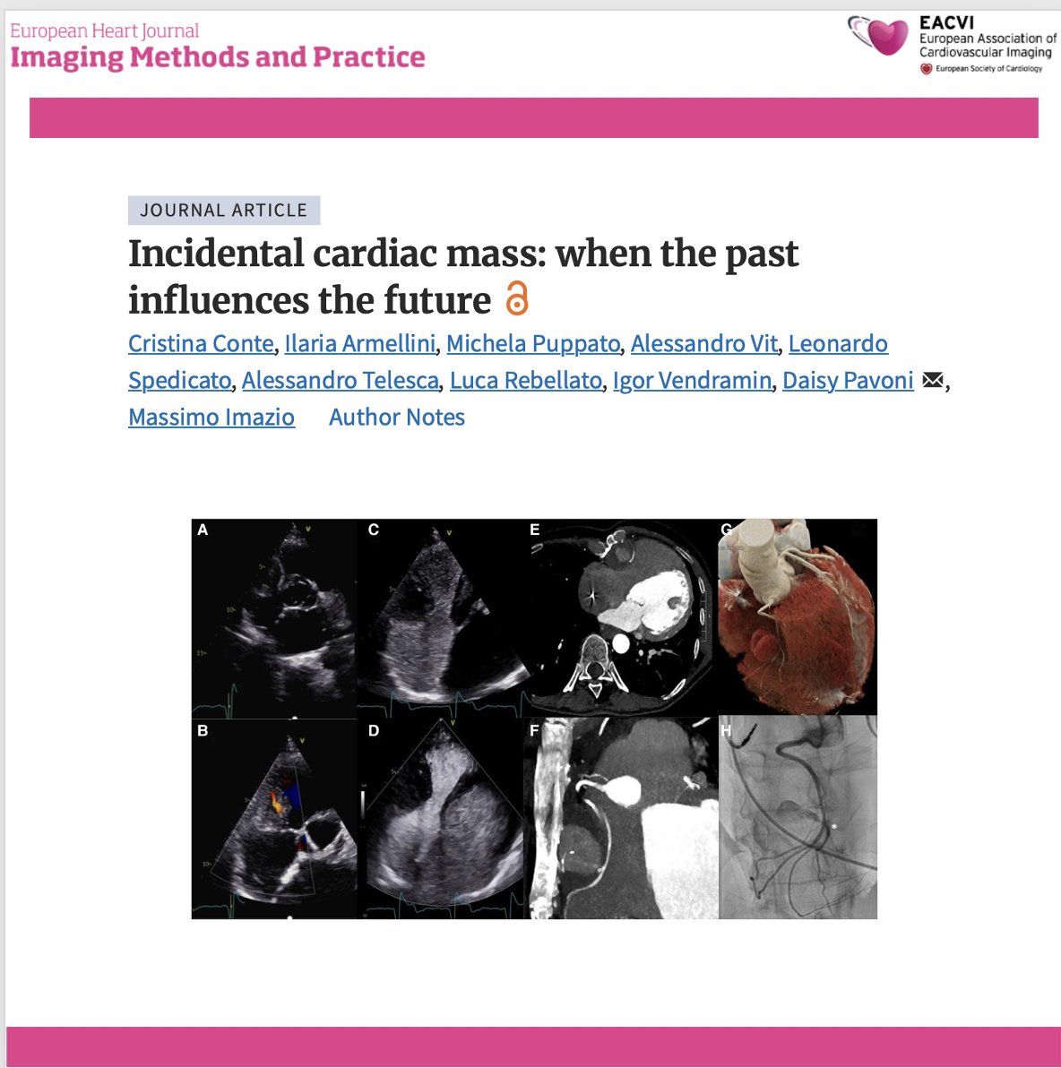
    🫀 When an incidental cardiac mass reveals a complication from the past… This striking multimodality imaging case describes a 53-year-old woman with previous surgical atrial septal defect closure in whom an incidental mediastinal mass was ultimately identified as a right coronary artery pseudoaneurysm with peripheral thrombosis—likely related to chronic erosion from nearby sternal wires. 🔍 Key findings: ✅ Transthoracic echocardiography detected a mass adjacent to the right atrioventricular groove, with systolic–diastolic flow and suspected circumferential thrombosis. ✅ Agitated saline excluded communication with the right heart, while contrast-enhanced ultrasound demonstrated delayed enhancement, suggesting a systemic arterial supply. ✅ Coronary CT angiography revealed marked sternal malalignment, close contact with the sternal wires, and kinking of the mid-to-distal right coronary artery. ✅ Invasive angiography confirmed active RCA perfusion, supporting the diagnosis of a coronary pseudoaneurysm. Because both percutaneous and surgical treatment were considered high risk, conservative management was chosen. 💡 Clinical takeaway: Incidental cardiac masses require more than anatomical description. Integrating echocardiography, contrast ultrasound, CT and invasive angiography can clarify vascular origin, define relationships with previous surgical material and guide complex management decisions. 📖 #CardioX #MultimodalityImaging #Echocardiography #CardiacCT #CoronaryPseudoaneurysm #CardiacMass #AdultCongenitalHeartDisease #CardiacSurgery #CardiacImaging #OpenScience
    Download
    011231.7K
  16. EHJ-IMP Editor-in-Chief✓@EHJIMPEiC
    🫀 Can routine cardiac PET provide prognostic information beyond myocardial perfusion? A study of 783 patients referred for rest–stress ¹⁵O-water PET shows that left ventricular hypertrophy can be quantified directly from routine, non-ECG-gated PET images—and may identify patients at increased cardiovascular risk. 🔍 Key findings: ✅ PET-derived LV mass index (LVMi) and interventricular septal wall thickness were significantly higher in patients with a previous diagnosis of heart failure. ✅ Among patients without known heart failure, both LVMi and wall thickness independently predicted major adverse cardiac events, defined as cardiovascular death or acute heart failure. ✅ The prognostic associations remained significant after adjustment for age, sex, and myocardial flow reserve. ✅ Measurements showed high test–retest repeatability, with no significant systematic bias in patients undergoing repeated PET examinations. 💡 Clinical takeaway: Routine ¹⁵O-water PET may offer more than perfusion assessment. Extracting LV hypertrophy metrics from the same examination could provide additional, reproducible prognostic information without further imaging or radiation exposure. 📖 #CardioX #NuclearCardiology #CardiacPET #PETImaging #LeftVentricularHypertrophy #HeartFailure #CoronaryArteryDisease #CardiacImaging #Cardiology #OpenScience
    Download
    1710635
  17. EHJ-IMP Editor-in-Chief✓@EHJIMPEiC
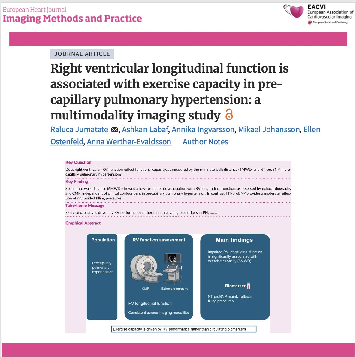
    🫀 What is the best imaging marker of exercise capacity in precapillary pulmonary hypertension? In precapillary pulmonary hypertension (PH), right ventricular (RV) adaptation determines prognosis. This multimodality imaging study shows that RV longitudinal function is more closely associated with exercise capacity than conventional biomarkers, highlighting its value for patient assessment. 🔍 Key findings: ✅ Impaired RV longitudinal function was independently associated with reduced exercise capacity, regardless of whether it was assessed by echocardiography or cardiac magnetic resonance. ✅ These associations remained significant after adjustment for clinical confounders, supporting RV longitudinal mechanics as a robust functional marker. ✅ In contrast, NT-proBNP was more strongly related to right-sided filling pressures than to functional capacity, suggesting it reflects a different aspect of disease severity. ✅ The findings reinforce the complementary role of multimodality imaging in evaluating RV performance beyond resting haemodynamics alone. 💡 Clinical takeaway: Resting haemodynamics and biomarkers tell only part of the story. Assessing RV longitudinal function may provide a more clinically meaningful estimate of functional limitation in patients with precapillary pulmonary hypertension and could improve risk stratification. 📖 Read the full article: #CardioX #PulmonaryHypertension #RightVentricle #Echocardiography #CardiacMRI #CardiacImaging #HeartFailure #Cardiology #MedTwitter #OpenScience
    Download
    0310672
  18. EHJ-IMP Editor-in-Chief✓@EHJIMPEiC
    🫀 What happens to the heart after transcatheter mitral valve repair with the PASCAL® system? Chronic mitral regurgitation leads to progressive cardiac remodelling, but how much reverse remodelling occurs after PASCAL® transcatheter edge-to-edge repair (TEER)? This study evaluates structural cardiac changes and their relationship with clinical outcomes following the procedure. 🔍 Key findings: ✅ Successful PASCAL® TEER was associated with favourable reverse cardiac remodelling, reflecting the haemodynamic benefit of reducing mitral regurgitation. ✅ Improvements in cardiac structure were accompanied by better clinical outcomes and functional status during follow-up. ✅ The study highlights the importance of serial imaging after TEER to monitor ventricular and atrial remodelling beyond procedural success. ✅ These findings further support the role of multimodality imaging in evaluating response to transcatheter mitral valve interventions and guiding long-term patient management. 💡 Clinical takeaway: The success of mitral TEER should not be judged solely by immediate MR reduction. Reverse cardiac remodelling is an important marker of sustained benefit, and follow-up imaging is essential to identify patients who derive the greatest long-term advantage. 📖 Read the full article: #CardioX #MitralRegurgitation #TEER #PASCAL #StructuralHeart #ValveDisease #CardiacImaging #Echocardiography #Cardiology #MedTwitter
    Download
    212271.8K